by CONNIE TABBERT
Editor
PEMBROKE — In a week’s time, the new MRI machine at Pembroke Regional Hospital will have been in operation for two weeks.
But, it’s only today that it’s officially available to all patients who require an MRI.
What is an MRI, you ask. An MRI (magnetic resonance imaging) scan is an imaging technicque that uses magnetism, radio waves and a computer to produce images of body structures.
The MRI scanner is a powerful magnet that creates a strong magnetic field, which aligns the protons of hydrogen atoms in the body. These atoms are then disrupted by a perpendicular pulse producing a response that is dependent on the specific tissue within the body the proton is located within.
The response produces a faint signal that is detected by the receiver portion of the MRI scanner. This information is processed by a computer, which produces the images.
Recently, Pierre Noel, Chief Executive Officer of Pembroke Regional Hospital, invited the media in to meet the experts dealing with the machine and to have a tour of the MRI suite.
“We are giving you the opportunity to come in, take pictures and learn about the MRI,” said Mr. Noel. “We want you to tell the story to our comunity about the MRI. Not only what it means to the community, but also what patients can expect when they come in.”
The experts at the event to explain the MRI, were radiologist technologists Lindsay Roosen and John Thomas, radiologists Dr. Fred Matzinger and Dr. Abram Choi, Bob Holmes, who is a fundraiser member for the hospital foundation and Sabine Mersmann, vice-president patient services.
Dr. Matzinger and Dr. Choi spoke about the technology while Ms. Roosen spoke about the machines and the safety aspects.
“You need to be very aware of some of the risks and the plans in place to deal with them,” Mr. Noel said.
He explained that the machinery and addition were funded 100 per cent by the community.
“When we turned on the machine on Monday, we started getting operating dollars for it,” he said. “When you deal with the government, you have to buy it and they will pay for the operating costs.”
The hospital recives funding to operate the MRI for eight hours a day, five days a week, he said.
“Over time that could change, but that’s what we’re being fund for,” he said.
There is also a new MRI in the Cornwall hospital, which also operates on limited hours, Mr. Noel said.
The Civic Campus of the Ottawa Hospital is the only place where the MRI operates 24 hours / seven days a week, Ms. Roosen said.
Mr. Noel explained that the Champlin LHIN (Local Health Integration Network), which operates from Hawkesbury to Barry’s Bay to just past Deep River, did an analysis, and the eight hours / five days a week, is what is required.
“For now, this is the capacity we need in our community, but there is projections that with an aging population and population growth, which is marginal in our area, we could be adding hours at some future point once proven we need more,” he said. “We’re content with that right now. If the situation proves we need more capacity to meet our demand, the opportunity is there to make a case for that.”
The hospital will receive about $800,000 a year, which pays for staffing and consumables, while physicians are remunerated through the typical remuneration mechanism, Mr. Noel said.
As for where the patients will come from, he said they could come from anywhere within the Champlin LHIN, as well as some from Quebec and the military base.
TECHNOLOGIST IS IN THE KNOW
Taking over from Mr. Noel, Ms. Roosen said all appointments are pre-booked and during the booking, there are several questions asked and advice given so the patient knows what to expect when they arrive.
When the patient arrives for their appointment, there is a more detailed screening form to fill out, because it’s very important there is no metal on or in the patient’s body, she said.
Emphasising the machine, she stressed, “The MRI is a giant magnet. It’s a 1.5 Tesla, which means it’s a super conducting magnet and it’s always on.
“If the power went off, the magnet would still be on on,” she said. “That’s why it’s very inportant we make sure patients are safe and metal free.”
The patients enter the waiting room and then are taken to a change room, which they cannot get to without special access, which means a technologist is with them from now until the MRI is completed, she said.
“We can’t have people not knowledgeable around the machine or walking by it,” Ms. Roosen said. “Anyone who has access is specially trained around the MRI and is aware of the dangers of the magnet always being on.”
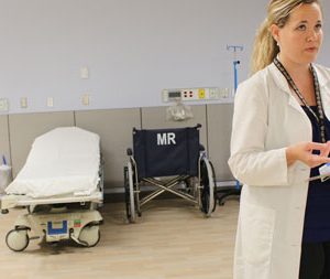
Once changed the patient is moved into Zone 2, which is the patient prep area. The patient is asked a series of questions, which they have most likely answered earlier, but they are reviewed again, for safety’s sake, she explained.
The patient is then whisked into Zone 3, which is the room just before entering the MRI (Zone 4).
“Wait times are precious, so there’s always someone in the machine and someone in the holding area,” Ms. Roosen said. “Our flow is fast and we try and see as many patients as we can in a day.”
Pointing to the floor, she noted there are different colour tile. Where it changes is how far the magnet can reach, she said.
“The magnet gets stronger as you get closer,” she added.
MRI times range from 20 minutes to an hour, depending on what the scanning is for, such as a knee or an elbow, brain scan or the abdomen, she said.
Ms. Roosen said depending on what body part or area is being scanned, there is various equipment, or coils, as they are professsionally called.
“These coils collect the image and converts it into the computer,” she said. “Every body part has a different (coil). There’s one for the foot, knee, abdomen, breast, pelvis, shoulder, head and neck.”
The body part to be scanned has to be in the centre of the magnet to get the best picture. So, if a person’s head is being scanned, that goes in first; the same for the feet. If it’s a hand, then the person lays on their stomach and the arm is stretch out ahead of them, she explained.
Ms. Roosen said, “The machine is very loud. It’s like a jackhammer, so every patient is given ear plugs and headphones. When the image is being taken, it takes about three to four minutes, and that’s when the machine is loud.
There are various safety measures for the patient at this time, she said. The technologist, who watches the patient from a glassed-in computer room, can hear the patient, see the patient and talk to the patient. The patient has a ball they can squeeze that can alert the technologist that something is wrong.
“At any time problems arise, such as a patient needs to come out, even if it’s becuause they don’t like it, they have this emegerncy ball. They squeeze it and it sets off an alarm for us. We stop the scan and ask if everything’s OK.”
Ms. Roosen added, “If we can see they are distressed from the back room, or if they are uncomfortable, or they want to talk to us, we can stop the scan and talk to them.”
While the patient is having the MRI, the radiologist technician watches the patient, not only thorugh a window but through a camera.
They can talk to the patient and the patient can talk back, she added.
Ms. Roosen said it’s important the patient does not move during the three to four minutes it takes to get one image. If the patient moves, the whole image is blurry, not just that single movement on film, she said.
If the patient moves, the image has to be re-taken, which means another three to four minutes of lying still for the same image.
“The MRI is not fast,” she said. “We can’t just take away the slice they moved on.”
Ms. Roosen said it’s the technologist who helps patients get through the whole thing.
“We’re coaxing them and telling them how good they’re doing,” she said.
She noted about three out of five patients are mildly claustrophic.
“Some can get through talking, or listening to music, or looking at mirrors,” she said. “They can also look out of the machine and see me the whole time and that seems to help patients. But then there are some patients who want to see nothing, so we give them blindfolds.
“It really depends on the person. We feel them out when we talk to them.”
EXCITING TIME
Dr. Matzinger said it’s been a “really exciting week for us here at the hospital.
“It’s the culmination of almost six years of fundraising by many people in the hospital and community to bring an MRI to Pembroke,” he said.
The staff at this hospital has always had one main objective and that is to “always provide high quality patient care close to home,” he said. “Thousands of patients from the Ottawa Valley and area travel to Ottawa every year for MRI scans. Now this is a new service we can provide.
“We are forutnate in having a state of the art piece of equipment,” Dr. Matzinger said.
The technologists began scanning patients and volunters on Sept. 14. By using volunteers, it ensured the technologists were able to practice scanning with this MRI before it was officially opened for general referrals, Mr. Noel said.
Dr. Matzinger said the hospital has “two excellent technolgoists. Image quality depnds not only on the MRI techinology, but also in having well trained technologists.”
Ms. Roosen grew up in Pembroke and has been working at the Civic Campus for about 10 years as an MRI technologist while Mr. Thomas has been training in MRI for about five years.
“We are lucky to have their expertise in the department,” Dr. Matzinger said.
He noted the MRI excells in imaging the brain, spine, joints and muscles, however, the hospital would never do without its CT scan as well.
“They have different uses, but they complement one another,” he said.
The CT is used more often in acute care, such as stroke, or a trauma occurring, he said.
As for reporting, Dr. Matzinger said there is a 24 to 48 hour turn-around period. The information is provided within that time frame to the physician who referred the patient and it is that physician who will inform the patient of the results, he explained.
While the two may overlap in their uses, Dr. Matzinger said sometimes one is better than the other for some diagnosis.
While there are private clinics where people can book their own MRIs, Dr. Matzinger noted only referrals from physicians will be accepted for patients at this MRI.
FUNDRAISING CONTINUES
Bob Holmes said the foundation assists the hospital in providing improved medical care for its patients.
The foundation receives and collects donations, as well as organizes and teams up with comunity partners to assist in various projects, such as dances, tournaments, lotteries, races, and other projects, both large and small, Mr. Holmes said.
He also spoke about the latest project, which is a health and home lottery that will help raise the last $230,000 of the original $4.5 million they started out to raise.
Tickets are available at the hospital, at the various sponsor locations and through volunteers who are out and about in the community.
Mr. Holmes encouraged everyone “to stop and talk to the volunters, find out what it’s all about and make a donation.”
Once the project funding is raised, Mr. Holmes said the foundation will not disappear.
“We will carry on in the future to assist Pembroke Regional Hospital as it tries to advance medicine.”
COMMUNITY EVENT
Mr. Pierre said it’s important the community who helped raise the money for the MRI see what they’ve purchased.
However, due to the small area, and the magnetism of the MRI, it really wouldn’t suit to have the public come to the hospital.
Instead, there will be a community information session at the Best Western Inn in Pembroke on Thursday, Oct. 29 from 5 to 7 p.m.
There will be formal remarks and a video presentation, he said.
As well, the early brid draw in the health and home lottery will be made at 6 p.m. followed by ligh refreshments.


![Kenopic/Smith Auction [Paid Ad]](https://whitewaternews.ca/wp-content/uploads/2018/10/advertising-100x75.jpeg)

